PERIO-PROSTHETIC GALLERY

Douglas H. Mahn, D.D.S.
Periodontics, Cosmetic Periodontal Surgery,
and Dental Implants
“Creating Beautiful and Healthy Smiles”
Call: 703-392-8844
Perio-Prosthetic Gallery
Periodontal - Restorative Smile Gallery:
A picture tells a thousand words. Please look through the Smile Gallery. All of the cases shown were performed by Dr. Mahn. Several were published in peer-reviewed dental journals and/or presented in lectures.
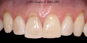
Connective Tissue Grafting Prior to Delivery of Cosmetic Restorations
Connective tissue grafting was performed by Dr. Mahn. Local dentists delivered the restorations.
Before:
After:
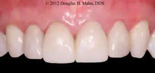
Published in Dentistry Today in Feb. 2012. Connective tissue grafting was performed to correct gingival recession and improve the stability of the gingival margin. Teeth were then treated with ceramic veneers.


Published in Dentistry Today in April 2015. Connective tissue grafting was performed to correct gingival recession and improve the stability of the gingival margin. Teeth were then treated with ceramic crowns.
Clinical Crown Lengthening
Clinical crown lengthening surgery was performed by Dr. Mahn. Local dentists delivered the restorations.
 of CL sx-Thornburg PreOp")
 of CL sx-Thornburg Post Op Final")
Severe tooth wear due to years of tooth grinding. Crown lengthening was required to provide sufficient tooth structure for restoration and to develop proper proportions. Teeth were restored with ceramic crowns.


The central incisors had a history of fracture. The patient was unhappy with the appearance of her teeth. The front teeth were treated with crown lengthening. Teeth were restored with ceramic crowns.
")
")
Published in Dentistry Today in Jan. 2011. Crown lengthening was required to close the space between the incisors while maintaining proper tooth proportions.


The top front teeth were fractured and repaired when the patient was a teenager. The patient was embarrassed by their appearance. Now in his mid-twenties, the patient was prepared to improve his smile. Clinical crown lengthening was required to help develop proper tooth proportions. Ceramic crowns were placed. The patient was very happy with the results of his treatment.


This patient was concerned about the short appearance of his anterior teeth and a history of tooth fracture. Clinical crown lengthening improved tooth-to-tooth proportions and enabled stable restorations to be placed. The patient was very pleased with the results of his treatment.


This patient had severely worn teeth from years of grinding. When he smiled, his teeth were not visible. Clinical crown lengthening was performed on all of his top teeth. This exposed enough tooth structure for proper and esthetic restorations to be placed. The patient was very pleased with the results of his treatment.


Published in Practical Periodontics and Aesthetic Dentistry in 2008. Crown lengthening was necessary to permit predictable and esthetic restoration of the top teeth. Ceramic crowns were placed on the top and bottom teeth.
Complex Periodontal-Restorative Reconstruction:
Crown lengthening, extractions, bone regeneration and dental implants was performed by Dr. Mahn. Local dentists delivered the restorations.


Mutilated dentition. Crown lengthening of top front teeth. Implants placed in posterior regions. Anterior teeth were restored with ceramic crowns. Fixed (non-removable) bridges replaced posterior teeth.

 of Copy of Hutchinson CL Final VERY BEST")
Severe tooth wear and erosion. Left central incisor had severe infection and was considered hopeless. This tooth was removed and the extraction site treated with bone regenerative techniques to prevent collapse of the remaining soft tissue architecture. The remaining teeth received crown lengthening surgery. A 3-unit fixed (non-removable) bridge was used to replace the left central incisor. The remaining anterior teeth received ceramic crowns.


This patient was unhappy with the misaligned and discolored appearance of his teeth. He did not want to have orthodontic treatment. Crown lengthening treatment established gingival margins that permitted esthetic restoration of his teeth. The front right lateral incisor was removed atraumatically to preserve the natural gingival contours. A 3-unit fixed (non-removable) bridge was used to replace the right lateral incisor. Ceramic crowns were placed the remaining anterior teeth.


This patient was very unhappy with the appearance of her smile and her front teeth. The space between her central incisors had developed over the last few years. Her left central and left lateral incisor had very severe periodontal infection and bone loss. These teeth were deemed hopeless. The left central and left lateral incisor were removed. These sites were treated with bone regenerative techniques to prevent collapse of the remaining soft tissue architecture and provide a foundation for esthetic replacement of her teeth. A fixed provisional bridge was placed the day of surgery. A final ceramic bridge was placed.


This patient was unhappy with the appearance of her smile and her front teeth. She had residual ridge resorption in areas where teeth were missing. The created a visible gap beneath the replacement tooth. The canine that had been orthodontically moved into the lateral incisor site developed gingival recession. Soft tissue grafting was used to improve the contours where teeth were missing and correct the gingival recession. Other techniques were used to transform the appearance of the canine to that of a lateral incisor. This was followed by tooth whitening and ceramic restorations.


This patient had dental implants placed by another doctor. The implant and tooth sites had a soft tissue architecture that did not permit proper restoration of these sites. I performed clinical crown lengthening on the tooth sites and ridge augmentation on the implant sites. The general dentist was then able to place highly esthetic crowns. The patient was thrilled with his new smile.
")
")
")
")
Published in The Journal of Esthetic and Restorative Dentistry in 2013. This patient had “canine substitution” orthodontics. She had her teeth moved so that her canine teeth replaced her congenitally missing lateral incisors. Unfortunately, she was unhappy with the appearance of her teeth and her gummy smile. To improve her smile, clinical crown lengthening surgery and bone recontouring was performed to permit esthetic restoration of her front teeth and elimination of the gummy smile. Ceramic restorations were placed on the front six teeth creating the appearance of natural incisors and canines with proper proportions.